Enzyme discovery may lead to better heart and stroke treatments
A Queen's University study sheds new light on the way one of our cell enzymes, implicated in causing tissue damage after heart attacks and strokes, is normally kept under control. Led by Biochemistry professor Peter Davies, the research team's discovery will be useful in developing new drug treatments that can aid recovery in stroke and heart disease, as well as lessen the effects of Alzheimer's and other neurologically degenerative diseases.
"This is particularly exciting because the enzyme structure we were seeking – and the way its inhibitor blocks activity without itself being damaged – have proved so elusive until now," says Dr. Davies, Canada Research Chair in Protein Engineering.
The team's findings will be published on-line in the international journal, Nature, on Thursday Nov. 20.
In remodeling proteins needed for cell growth and movement, our cells use the enzyme calpain to break off pieces from other proteins. Calpain is activated when the cell releases short bursts of calcium.
During heart attacks or strokes, however, blood supply to cells is interrupted. When the blockage is re-opened, the influx of blood causes calcium levels in the cell to become dangerously high, and the calpain activity to increase. The result is significant damage to tissues. "While you want the enzyme to switch on and off, you don't want it to go out of control," says Biochemistry research associate Rob Campbell, a member of the Queen's team.
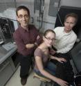
The study shows how another protein, calpastatin, binds and blocks calpain once it has been activated by calcium. Dr. Campbell, an x-ray crystallographer, and PhD student Rachel Hanna were able to determine the structure of the calcium-bound calpain and discover how calpastatin can inhibit calpain without being cut and destroyed in the process. That information will be useful in designing drugs to protect against the damage caused by over-activation of calpain.
Because the crystals grown in the lab at Queen's were too small to be used for X-ray diffraction data collection on the university's diffractometer, Dr. Campbell and Ms Hanna booked time on the National Synchrotron Light Source at Brookhaven Long Island (operated by the U.S. Department of Energy).
Working in shifts around the clock, they collected the required data during the first nine of their 48 allotted hours. After another hour, "We knew we had the structure solved," Ms Hanna recalls. "It was really exciting. We immediately sent an e-mail to Peter to say: 'We did it!'"
Source: Queen's University
Other sources
- Enzyme Discovery May Lead To Better Heart And Stroke Treatmentsfrom Science DailyThu, 20 Nov 2008, 0:14:32 UTC
- Enzyme discovery may lead to better heart and stroke treatmentsfrom PhysorgWed, 19 Nov 2008, 18:35:45 UTC
